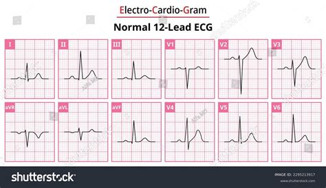
normal ecg 12 lead|ECG interpretation: Characteristics of the normal ECG : Pilipinas ECG Interpretation: definitions, criteria, and characteristics of the normal ECG waves, intervals, durations and rhythm. This is arguably one of the most important chapters throughout this course. At the heart of ECG interpretation lies the .
striort reclamações El pronóstico y sugerencia de apuesta para Palmeiras vs Tombense, con fecha 12 de abril de 2023, en el análisis escrito por los editores de Pronósticos Deportivos, va a: Palmeiras ganará en el medio tiempo ⇒ apuesta disponible en Bet365. El Palmeiras vs Tombense del 12 de abril de 2023 se jugará en São Paulo, Allianz Parque.

normal ecg 12 lead,Learn how to diagnose a normal adult 12-lead ECG by excluding any recognised abnormality. See the normal ranges and causes of P wave, PR interval, QRS complex, QT interval, ST segment and T wave.12-lead ECG library, Sinus Bradycardia. A 55 year old man with 4 hours of .The 12 lead ECG library - ecglibrary.com. A collection of electrocardiograms. . The .If the axis is in the "left" quadrant take your second glance at lead II. both I and aVF . ECG features of normal sinus rhythm. Regular rhythm at a rate of 60-100 bpm (or age-appropriate rate in children) Each QRS complex is preceded by a normal P wave. Normal P wave axis: P waves upright in leads I and II, .
normal ecg 12 lead ECG interpretation: Characteristics of the normal ECG ECG Interpretation: definitions, criteria, and characteristics of the normal ECG waves, intervals, durations and rhythm. This is arguably one of the most important chapters throughout this course. At the heart of ECG interpretation lies the .Is the electrical axis normal? Electrical axis is assessed in limb leads and should be between –30° to 90°. Common findings. Wide QRS complex (QRS duration ≥0.12 s): Left bundle .normal ecg 12 lead Normal findings include: * Normal sinus rhythm. The rhythm is regular, the rate is 80 bpm, and there is a P wave before every QRS complex. The P waves all look alike in each lead, and they are upright in the inferior .
Normal: 60-100 bpm. Tachycardia: > 100 bpm. Bradycardia: < 60 bpm. Regular heart rhythm. If a patient has a regular heart rhythm, their heart rate can be calculated using the following method: Count the number of large .•Describe how the 12 lead ECG portrays the electrical activity of the heart •Differentiate between ischemia, injury and infarction
An ECG lead is a graphical representation of the heart’s electrical activity which is calculated by analysing data from several ECG electrodes. A 12-lead ECG records 12 leads, producing 12 separate graphs on a piece of ECG .
The Standard 12 Lead ECG. The standard 12-lead electrocardiogram is a representation of the heart's electrical activity recorded from electrodes on the body surface. This section describes the basic components of the ECG and .
The 12-lead ECG misleadingly only has 10 electrodes (sometimes also called leads but to avoid confusion we will refer to them as electrodes). The leads can be thought of as taking a picture of the heart’s electrical activity from 12 . Typical ECG findings for normal cardiac axis: Lead II has the most positive deflection compared to leads I and III. Normal Cardiac Axis Right axis deviation. . How to record a 12-lead ECG: an OSCE guide to recording .Numerous ECG lead systems and constellations of leads have been tested but the standard 12-lead ECG is still the most used and the most important lead system to master. The 12-lead ECG offers outstanding possibilities to .Normal 12-lead ECG Tracing | Learn the Heart - Healio A normal ECG trace includes a P wave, a QRS complex and a T wave. A standard 12-lead ECG includes bipolar limb leads, unipolar limb leads and chest leads.

The 12-lead ECG misleadingly only has 10 electrodes (sometimes also called leads but to avoid confusion we will refer to them as electrodes). . Normal duration of ECG segments: PR interval: 0.12 – 0.2 secs (3-5 small squares) QRS: <0.12 secs (3 small squares) QTc: 0.38 – 0.42 secs .A normal ECG is illustrated above. Note that the heart is beating in a regular sinus rhythm between 60 - 100 beats per minute (specifically 82 bpm). All the important intervals on this recording are within normal ranges. 1. P wave: upright in leads I, aVF and V3 - V6; normal duration of less than or equal to 0.11 seconds
The goal of the electrocardiogram interpretation is to determine whether the ECG waves and intervals are normal or pathological. Electrical signal interpretation gives a good approximation of heart pathology. A standard 12 lead ECG is shown in [Figure 1]. The best way to interpret an ECG is to read it systematically: RateECG Diagnosis: ECG Limb Lead Reversal. ECG Limb Lead Reversal: ECG Diagnosis: Upper Limb Lead reversal. Upper Limb Lead reversal: ECG Diagnosis: Block: Left Anterior Fascicular Block (LAFB) LAFB, left anterior hemiblock, left anterior hemi-block: ECG Diagnosis: Left Axis Deviation. Left Axis Deviation, LAD: ECG Diagnosis: Block: Left Bundle .Normal adult 12-lead ECG. The diagnosis of the normal electrocardiogram is made by excluding any recognised abnormality. It's description is therefore quite lengthy. normal sinus rhythm each P wave is followed by a QRS P waves normal for the subject P wave rate 60 - 100 bpm with <10% variation rate <60 = sinus bradycardia1. The Standard 12 Lead ECG. The standard 12-lead electrocardiogram is a representation of the heart's electrical activity recorded from electrodes on the body surface. This section describes the basic components of the ECG and the lead system used to record the ECG tracings. Topics for study: ECG Waves and Intervals; Spatial Orientation of the . Of course, there are many variations in ECGs considered to be normal. Once a student recognizes the features of the normal ECG, it becomes possible to recognize “abnormal” and then learn the clinical ramifications of . Thomas Lewis developed and described (1913) his lead configuration to magnify atrial oscillations present during atrial fibrillation. When fibrillation is present and the electrodes lie in the vicinity of the right auricle . An electrocardiogram (EKG or ECG) is a test that measures the electrical activity of the heartbeat. The American Heart Association explains an electrocardiogram (EKG or ECG) is a test that measures the electrical activity of the heartbeat. . A normal heartbeat on ECG will show the rate and rhythm of the contractions in the upper and lower .

ECG interpretation clearly illustrated by Professor Roger Seheult, MD. This is video 1 of the MedCram ECG online course: https://www.medcram.com/courses/ekg-.ECG interpretation: Characteristics of the normal ECG ECG interpretation clearly illustrated by Professor Roger Seheult, MD. This is video 1 of the MedCram ECG online course: https://www.medcram.com/courses/ekg-. Method 2: Three Lead analysis – (Lead I, Lead II and aVF) Next we add in Lead II to the analysis of Lead I and aVF . A positive QRS in Lead I puts the axis in roughly the same direction as lead I.; A positive QRS in Lead II similarly aligns the axis with lead II.; We can then combine both coloured areas and the area of overlap determines the axis.
Pathophysiology. At birth, the right ventricle is larger and thicker than the left ventricle, reflecting greater physiological stresses placed upon it in utero (i.e. pumping blood through the relatively high-resistance pulmonary circulation).. This produces an ECG picture reflecting that of a right ventricular strain pattern in adults:. T-wave inversions in V1-3 In general, an EKG collects information from 12 different areas of the heart. The fact that electrical signals generated in the heart do not travel evenly over the skin is used in the case of a 12-lead EKG. Comparing the signals from the 12 different leads, the doctors can point out the exact location of a problem in the heart. An EKG test is a fast, painless way for your healthcare provider to learn about the frequency and strength of the electrical impulses that control your heartbeats. . Wear a shirt that you can remove easily to place the leads on your chest. What to expect on the date of the EKG test. A healthcare provider will attach 12 electrodes with .
normal ecg 12 lead|ECG interpretation: Characteristics of the normal ECG
PH0 · Understanding an ECG
PH1 · Reference (normal) values for ECG (electrocardiography)
PH2 · Normal Sinus Rhythm • LITFL Medical Blog • ECG
PH3 · Normal 12
PH4 · How to Read an ECG
PH5 · ECGlibrary.com: Normal adult 12
PH6 · ECG interpretation: Characteristics of the normal ECG
PH7 · ECG (EKG) Interpretation
PH8 · 12 Lead ECG Interpretation
PH9 · 1. The Standard 12 Lead ECG