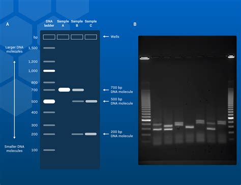
visualizing gel electrophoresis results|Gel electrophoresis: Visualising and interpreting the results : Manila Agarose, produced from seaweed, is a polysaccharide agar. During polymerization, agarose polymers link non-covalently and form a network of bundles. This network consists of pores with molecular filtering properties. Conceptual Rendering of Agarose Gel at a Microscopic Level. DNA separation occurs . Tingnan ang higit pa We would like to show you a description here but the site won’t allow us.We would like to show you a description here but the site won’t allow us.

visualizing gel electrophoresis results,Agarose, produced from seaweed, is a polysaccharide agar. During polymerization, agarose polymers link non-covalently and form a network of bundles. This network consists of pores with molecular filtering properties. Conceptual Rendering of Agarose Gel at a Microscopic Level. DNA separation occurs . Tingnan ang higit pa

The gel electrophoresis conditions (including the presence of ethidium bromide, gel concentrations, electric field strength, . Tingnan ang higit pa
Agarose Products Agarose LE (Molecular Biology Grade) (Catalog No. A-201) High Resolution Agarose (For Nucleotides < 1kb) (Catalog No. A-202) Low Melt Agarose . Tingnan ang higit pa
Dis 10, 2018 — Wrapping up: How to read gel electrophoresis results? First, make clear if a gel contains any results or not. For that, put the gel carefully under the UV light and see .
Hun 13, 2023 — Gel electrophoresis is an essential molecular biology technique used in biotechnology labs to separate and analyze nucleic acids (DNA fragments, RNA, and plasmids) and proteins based on their .
Visualizing the Results with Electrophoresis. Once a PCR reaction has been completed, we need to be able to see the results. To do this, a sample of the PCR mixture is loaded into an agarose gel for .Nob 20, 2007 — Gel electrophoresis is used to separate macromolecules like DNA, RNA and proteins. DNA fragments are separated according to their size. Proteins can be separated according to their size and their .The five main steps in nucleic acid gel electrophoresis are gel preparation, sample and ladder preparation, electrophoretic run, sample visualization, and gel documentation. Click on the tiles to deep dive into each step. 1. .Gel electrophoresis is used to characterize one of the most basic properties - molecular mass - of both polynucleotides and polypeptides. Gel electrophoresis can also be used to determine: (1) the purity of these .Step 1: Record clear digital images of your electrophoresis gel. The first step in assessing electrophoresis gels should be to record a good digital image of the gel. Digital images allow closer inspection of very small .How to Read a Gel Image – edias-project. ~We use gel electrophoresis to verify our success of DNA extraction and PCR amplification~ DNA Ladder & Negative Controls. It is important to load the gel with a ladder to be used .DNA gel electrophoresis is a technique used for the detection and separation of DNA molecules. An electric field is applied to a gel matrix comprised of agarose, and within the gel, charge particles will migrate .
Khanmigo is now free for all US educators! Plan lessons, develop exit tickets, and so much more with our AI teaching assistant.Agarose gel electrophoresis is a method of choice for large molecule separation over 1 million Da. Acrylamide cannot be used for this purpose, because it remains liquid at the concentration required for the appropriate separation of high-molecular-weight analytes. The movement of molecules through an agarose gel is dependent on the size and charge of .
Gel electrophoresis of nucleic acids is an analytical technique to separate DNA or RNA fragments by . Capillary electrophoresis results are typically displayed in a trace view called an . By running DNA through an EtBr-treated gel and visualizing it with UV light, any band containing more than ~20 ng DNA becomes distinctly visible. .When visualizing this PCR reaction, two bands should appear in the same lane if Wolbachia is present, and only one band . To interpret gel electrophoresis results, first ensure that all controls are correct. The DNA ladder, (+) Arthropod control, (-) Arthropod control, and (+) DNA control should produce bands of expected size, whereas the .Possible causes for smearing in gel electrophoresis Recommendations to minimize smearing in gel electrophoresis; Thick gels: Keep the gel thickness around 3–4 mm when casting horizontal agarose gels. Gels thicker than 5 mm may result in band diffusion during electrophoresis. Poorly formed wells: Use clean gel combs when casting gels.Visualizing Separated DNA Fragments. 6:27 . DNA Gel Electrophoresis is a technique used to separate and identify DNA fragments based on size. . Here you see an agarose gel electrophoresis result after separating PCR products. The DNA fragments loaded into the gel are visible as clearly defined bands. The DNA standard or ladder should be .
Visualizing gel electrophoresis results After a gel run is complete, the samples must be visualized. Since nucleic acids are not visible under ambient light, special detection methods are required for visualization. In the tabbed section below, Tables .Gel electrophoresis works because DNA is negatively charged, due to the presence of phosphate groups in its backbone. Samples of PCR product are loaded into wells in the gel near the negative node. Due the the electric current across the buffer and gel matrix, DNA fragments migrate towards the positive node and separate by size.visualizing gel electrophoresis results Gel electrophoresis: Visualising and interpreting the resultsDis 15, 2022 — Figure 8. Electrophoresis: A gel electrophoresis set-up with agarose gel with DNA and loading dye on the left and the power supply on the right. Image Source: Michael, CC BY 2.0, via Wikimedia Commons and U. S. Department of Agriculture, CC BY 2.0, via Wikimedia Commons. Figure 8 shows a picture of a gel electrophoresis gel .Gel electrophoresis: Visualising and interpreting the resultsVisualizing gel electrophoresis results After a gel run is complete, the samples must be visualized. Since nucleic acids are not visible under ambient light, special detection methods are required for visualization. In the tabbed section below, Tables .Gel electrophoresis of proteins is a laboratory technique that allows the separation and analysis of proteins based on their size, shape, and charge. In this module, you will learn the principles and applications of gel .Abr 20, 2012 — Agarose gel electrophoresis is the most effective way of separating DNA fragments of varying sizes ranging from 100 bp to 25 kb 1.Agarose is isolated from the seaweed genera Gelidium and Gracilaria, and consists of repeated agarobiose (L- and D-galactose) subunits 2.During gelation, agarose polymers associate non-covalently and .
You will use gel electrophoresis to separate and visualize DNA fragments. Loading the gel. Join Dr. One in the molecular biology lab. The DNA samples from the crime scene and two suspects have already been .Hun 1, 2021 — 3.1 Protocol Native Electrophoresis. 1. Prepare a 3–8% or 4–10% acrylamide/bisacrylamide (AAB) gradients for hrCN, BN, and hybrid gels, respectively. Table 1 summarizes the quantities of buffer, AAB, H 2 O, glycerol, ammonium persulfate (APS), and tetramethylethylenediamine (TEMED) used for a mini-gel (size of the gel: 85 .
visualizing gel electrophoresis resultsHun 1, 2021 — 3.1 Protocol Native Electrophoresis. 1. Prepare a 3–8% or 4–10% acrylamide/bisacrylamide (AAB) gradients for hrCN, BN, and hybrid gels, respectively. Table 1 summarizes the quantities of buffer, AAB, H 2 O, glycerol, ammonium persulfate (APS), and tetramethylethylenediamine (TEMED) used for a mini-gel (size of the gel: 85 .the bottom of the gel well, instead of diffusing in the buff er. 9 Once solidied, place the agar ose gel into the gel box (electrophoresis unit). 10 Fill gel box with 1xTAE (or TBE) until the gel is co vered. Note *Pro-Tip* Remember, if you added EtBr to your gel, add some t o the buffer as well. EtBr is positiv elyDis 10, 2018 — PCR gel electrophoresis result from image 1. The image is captured under the UV transilluminator instead of the gel doc system to show you the effect of EtBr on the gel electrophoresis results. Here due to the re-use of a gel as well as the buffer, the EtBr is not properly spread into the gel.
Gold Biotechnology (U.S. Registration No 3,257,927) and Goldbio (U.S. Registration No 3,257,926) are registered trademarks of Gold Biotechnology, Inc.
Visualizing gel electrophoresis results After a gel run is complete, the samples must be visualized. Since nucleic acids are not visible under ambient light, special detection methods are required for visualization. In the tabbed section below, Tables .
visualizing gel electrophoresis results|Gel electrophoresis: Visualising and interpreting the results
PH0 · Steps in Nucleic Acid Gel Electrophoresis
PH1 · Interpreting Electrophoresis Gels with Bento Lab
PH2 · How to Read, Interpret and Analyze Gel Electrophoresis Results?
PH3 · How to Read a Gel Image – edias
PH4 · How to Interpret DNA Gel Electrophoresis Results
PH5 · How To Read & Interpret Gel Electrophoresis
PH6 · Gel electrophoresis: Visualising and interpreting the results
PH7 · DNA Gel Electrophoresis: Concept, Procedure, and
PH8 · 3.1: Gel Electrophoresis
PH9 · 1.4: PCR and Gel Electrophoresis