what is nonspecific st and t wave abnormality|ECG T Wave : iloilo Discover the 68 causes of T wave and ST segment abnormalities in ECG interpretation with Learn the Heart on Healio.
Current exchange rate US DOLLAR (USD) to PHILIPPINES PESO (PHP) including currency converter, buying & selling rate and historical conversion chart.

what is nonspecific st and t wave abnormality,Mar 4, 2020 — Isolated nonspecific ST-segment and T-wave abnormalities (NSSTTAs) are common findings on the resting ECG in asymptomatic individuals. 1 The prevalence of these isolated minor abnormalities as defined by Minnesota ECG classification ranges .Mü °í›8 B˘ww+qœÕü J/$ È}XÎ p71ÀÆ©¡Ü ´ `CÒhàò §]t ”´¸– &V óí\‘ hg+¶Ê .The most common ECG changes are nonspecific ST-segment and T-wave .May 19, 2023 — What are non-specific T wave changes? They occur in about 1% of patients and include T wave flattening and T wave inversion with no other changes necessarily present. Causes include (1): .
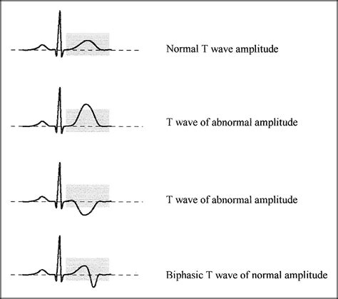
Ene 10, 2013 — Although very common, nonspecific ST-T (NSST-T) wave changes on ECG are often misunderstood, poorly explained to patients, or prematurely dismissed by .what is nonspecific st and t wave abnormalityDiscover the 68 causes of T wave and ST segment abnormalities in ECG interpretation with Learn the Heart on Healio.what is nonspecific st and t wave abnormality ECG T Wave Discover the 68 causes of T wave and ST segment abnormalities in ECG interpretation with Learn the Heart on Healio.Ene 9, 2023 — The most common ECG changes are nonspecific ST-segment and T-wave abnormalities, which may occur because of focal myocardial injury or ischemia caused by the metastatic tumor. In some .Set 22, 2015 — Changes in the ST segment and T waves can be early markers of an underlying cardiovascular disease, and even minor ST‐T abnormalities have predicted .NONSPECIFIC ST-T-WAVE CHANGES. Nonspecific ST-T-wave changes are very common and may be seen in any lead of the electrocardiogram. The changes may be seen in .In previous studies, isolated non-specific ST-segment and T-wave abnormalities (NSSTTAs), a common finding on electrocardiograms (ECGs), were associated with .The T-wave may diminish in amplitude (flat T-waves), become negative (T-wave inversion) or even increase markedly in amplitude (hyperacute T-wave). Which of these ST-T changes occur depends on the localization, .Dis 22, 2022 — Tall T-waves (hyper-acute T waves) can be an early sign of ST-elevation myocardial infarction. The morphology of the T waves can begin to broaden and peak within 30 minutes of complete coronary .
Q: I am a 41 years old man and I underwent a routine ECG and the report showed sinus rhythm, left axis, non-specific ST-T abnormality (elevated).Otherwise it was a normal ECG. What does it mean? A:ST segment and T wave are ECG terminologies and these are arbitrary names given to certain segments of the tracings of the ECG.ST-T wave .
Mar 31, 2017 — Nonspecific ST-segment and T-wave (ST-T) changes represent one of the most prevalent electrocardiographic abnormalities in hypertensive patients. However, a limited number of studies have investigated the association between nonspecific ST-T changes and unsatisfactory blood pressure (BP) control in adults with hypertension. .Ene 30, 2018 — BackgroundT‐wave abnormalities are common during the acute phase of non‐ST‐segment elevation acute coronary syndromes, but mechanisms underlying their occurrence are unclear. We hypothesized .
Dis 8, 2008 — Minor ECG abnormalities, especially minor nonspecific ST-segment and T-wave abnormalities (NSSTTAs), are common in asymptomatic individuals and often occur in the absence of other ECG abnormalities. 1–4 Isolated minor NSSTTAs generally represent very minor or upsloping ST-segment depression and flat or minimally inverted .

Nonspecific repolarization abnormalities in the ST-T segment are prevalent in patients with prehospital chest pain. Patients with such ECG patterns are more likely to be admitted to the hospital, are more likely to have a cardiac-related etiology of chest pain, have on average 1-day increased length of stay, and have two-fold increased risk of .
Hun 4, 2020 — As the American Heart Association (AHA) describes it, the right and left atria (the upper chambers, or ventricles) make a wave called a “P wave,” the bottom right and left chambers make a wave .Abr 25, 2024 — T wave: This shows the electrical activity involved in the heart’s ventricular repolarization. This means it shows the electrical reset of the heart as it prepares for the next cardiac cycle.
Ago 15, 2012 — Most clinicians regard isolated, minor, or nonspecific ST-segment and T-wave (NS-STT) abnormalities to be incidental, often transient, and benign findings in asymptomatic patients. We sought to evaluate whether isolated NS-STT abnormalities on routine electrocardiograms (ECGs) are associated with in .Ene 30, 2018 — BackgroundT‐wave abnormalities are common during the acute phase of non‐ST‐segment elevation acute coronary syndromes, but mechanisms underlying their occurrence are unclear. We hypothesized that T‐wave abnormalities in the presentation of non‐ST‐segment elevation acute coronary syndromes correspond to the presence of .

The initial electrocardiographic analysis of an electrocardiogram was based on visual pattern recognition and interval measurements. Indeed, when the first human electrocardiograms were recorded on photographic plates, only the crudest interval measurements were possible. In 1887, Waller1 showed that the changes in the electrical .Peb 9, 2023 — Hi @ih60, it sounds like you've received your ECG results before having a chance to review them with your cardiologist.ST- and T-wave changes can mean many different things and the interpretation of the findings, needs the expertise of a medical professional who is familiar with your medical history.
The transition from ST segment to T-wave is smooth, and not abrupt. ST segment deviation (elevation, depression) is measured as the height difference (in millimeters) between the J point and the baseline (the PR .Dis 22, 2022 — Abnormal T-wave Etiology. Abnormalities in the T-wave may represent variations of normal cardiac electrophysiology or signs of pathology. Tall T-waves (hyper-acute T waves) can be an early sign of .
As noted above, the transition from the ST segment to the T-wave should be smooth. The T-wave is normally slightly asymmetric since its downslope (second half) is steeper than its upslope (first half). Women have a more symmetrical T-wave, a more distinct transition from ST segment to T-wave and lower T-wave amplitude.Peb 10, 1999 — Nonspecific abnormalities are frequently observed in tracings of persons without clinical signs of heart disease. The most common nonspecific findings, ST segment or T-wave abnormalities or both (ST-T abnormalities), can be disquieting hints of latent abnormality that the physician may not be able to confirm or completely dismiss.Dis 15, 2017 — Nonspecific ST-segment and T-wave changes in symptomatic patients should not be considered normal and should prompt further evaluation for a cardiac cause. C 25
ECG T Wave A study was made of 1,000 consecutive adult in-patient electrocardiograms to determine the possibility of making a more precise diagnosis than "nonspecific ST and T-wave changes." More than 50 per cent (209) of the 410 abnormal electrocardiograms (exclusive of arrhythmias) were characterized by nonspecific depression of ST segment or T wave .Introduction. Isolated non-specific ST-segment and T-wave abnormalities (NSSTTAs) are common findings on the resting electrocardiogram (ECG) in asymptomatic individuals. 1 The prevalence of these isolated minor abnormalities as defined by Minnesota ECG classification ranges from 3.6% to 10.3% and they are consistently more prevalent .
what is nonspecific st and t wave abnormality|ECG T Wave
PH0 · What Is the Truth Behind Abnormal ECG Changes?
PH1 · The Non
PH2 · Electrocardiographic T Wave Abnormalities and the Risk of
PH3 · Electrocardiographic ST
PH4 · ECG tutorial: ST and T wave changes
PH5 · ECG in myocardial ischemia: ischemic changes in the
PH6 · ECG T Wave
PH7 · ECG Learning Center
PH8 · Communicating Concerns About Nonspecific Changes on ECG
PH9 · 68 causes of T wave, ST segment abnormalities